Last updated: September 25, 2026
Joints bear burdens silently until they begin to protest. Every step down a flight of stairs or a simple walk across a room places forces several times our body weight directly onto the knee cartilage. Underneath the kneecap and between the femur and tibia lies a specialized fluid that dampens these forces. Over decades, this internal cushioning system degrades. The biological fluids lose their thickness, and bone surfaces experience higher friction during everyday joint stress.
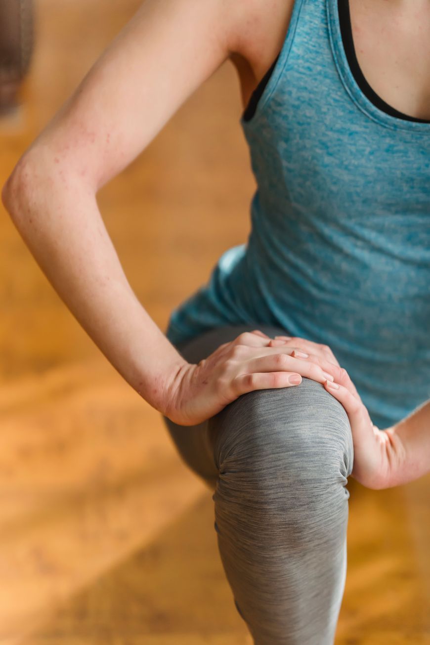
Age alters nearly every tissue in the human body, but the degradation of weight-bearing joint mechanics carries painful consequences. People often notice stiffness first thing in the morning or a dull ache after prolonged standing. These early signals point toward a deeper chemical shift inside the capsule. The biological components responsible for keeping movement smooth begin to break down, accelerating knee osteoarthritis progression if left unchecked.
The Chemistry of Fluid Dynamics
Healthy movement relies on a thick substance that coats the articular cartilage. This biological fluid behaves strangely under pressure. When a sudden impact occurs, the fluid thickens instantly to absorb the shock, protecting the underlying bone. During slow movements, it thins out to act as a lubricant. This dual nature comes down to complex molecular chains floating in water.
A significant reduction of hyaluronic acid concentration with age disrupts this delicate balance. Without enough of these long-chain molecules, synovial fluid viscoelasticity drops sharply. Researchers measure this property to understand how well a joint handles stress. When the fluid loses its structure, cartilage-on-cartilage friction increases. A 2021 clinical review published in the Journal of Orthopaedic Research noted that elderly joints exhibit a fifty percent reduction in macromolecular hyaluronan chain length compared to younger cohorts. That structural loss translates directly to lost protection.
The Role of Synthetic Lubrication
Scientists realized decades ago that replacing the worn fluid inside the capsule could alter the trajectory of degenerative joint disease. This therapeutic approach, known as viscosupplementation, introduces manufactured or purified lubricating gels directly into the joint space. The goal remains simple: restore joint cushioning and buy time for aging tissues.
Modern solutions often rely on cross-linked hyaluronan to mimic or exceed the resilience of natural biological fluids. One prominent example in this category is Synvisc, a high-molecular-weight fluid replacement derived from avian sources that is designed to restore viscoelastic properties in compromised joints. If you are a clinician looking to order Synvisc injections for your practice, you will find that these preparations fill the joint cavity, coating the cartilage surfaces and absorbing loads that would otherwise damage the bone. Medical professionals administer Synvisc to provide reliable mechanical stability when conservative measures fail.
The introduction of Synvisc alters the biomechanical environment by temporarily substituting for depleted native fluid. Because Synvisc mimics healthy high-molecular-weight hyaluronic acid, it temporarily halts the grinding friction that worsens cartilage wear.
Laboratory Insights Into Joint Mechanics
Laboratory experiments and clinical trials shed light on how these treatments function at a microscopic level. When researchers test synovial fluid substitutes in artificial joint simulators, they observe an immediate drop in friction coefficients. The injected material coats the micro-asperities of the cartilage, creating a microscopic barrier.
Studies tracking knee joint lubrication over six-month periods show that high-molecular-weight gels remain active within the capsule long after the initial procedure. They stimulate resident cells to produce healthier endogenous fluids. The mechanics improve not through physical presence alone, but through biological signaling.
The Progression of Cartilage Wear
Joint degeneration rarely happens overnight. Years of accumulated micro-trauma wear away the smooth outer layer of articular cartilage. As the protective surface thins, the underlying bone reacts by forming small growths known as osteophytes. These bony spurs alter the natural alignment of the joint, causing further discomfort during movement.
- Cartilage gradually loses its smooth outer texture and elasticity.
- Bone surfaces begin to experience direct contact under heavy loads.
- Inflammatory chemicals accumulate inside the synovial capsule.
These physical changes create a feedback loop of irritation and further fluid breakdown. Breaking this cycle requires more than simple pain management; it demands a restoration of the physical properties lost to time.
Restoring Shock Absorption
Treating aging knees effectively means looking closely at how the body handles physical impact. Shock absorption in the knee depends entirely on the health of the surrounding capsule. Without adequate padding, every step sends a sharp shock wave up the tibia and into the hip.
Physicians and researchers continue to evaluate different methods for restoring lost volume. Patients frequently ask about options that go beyond standard anti-inflammatory medications. They want solutions that address the root mechanical failure. By introducing targeted substances into the joint cavity, medical professionals can recreate the damping effect lost during the natural aging process.
Preserving Mobility Over Time
Living with aging knees requires an understanding of how daily habits influence joint fluid replenishment. Low-impact movement stimulates the remaining synovial lining to secrete fresh fluid, keeping the internal environment active. Complete rest often backfires, causing the fluid to lose its remaining viscosity.
Physical therapy protocols focus on building surrounding muscle strength to offload the joint, while treatments targeting shock absorption in the knee handle the internal friction. Balancing mechanical load with chemical restoration remains the primary objective in modern orthopedic management.
Cellular turnover within the joint capsule slows down considerably as decades pass. Chondrocytes, the specialized cells responsible for maintaining the extracellular matrix, become less responsive to mechanical and chemical stimuli. This sluggish metabolic rate means that damaged cartilage repairs itself at a fraction of its former speed.
Biochemical analysis of synovial fluid reveals a direct correlation between decreased volume and rising discomfort levels. When the body stops producing sufficient amounts of high-molecular-weight polymers, the remaining liquid becomes watery and less capable of energy dissipation. The joint loses its natural shock absorber.
Pathological changes in the subchondral bone compound this failure. As the cartilage wears thin, the bone beneath undergoes remodeling, becoming denser and stiffer. This loss of compliance means the entire skeletal structure absorbs harsh impacts rather than the fluid-filled cavity.
Therapeutic interventions must therefore address both the fluid medium and the hard tissue interface. Injectable gels serve as artificial placeholders, restoring the spacing and cushioning necessary to prevent bone-on-bone grinding. When administered correctly, these viscous solutions reintroduce the missing resistance against compressive loads.
Clinical data suggests that repeated supplementation can maintain functional mobility for extended periods. Patients report sustained improvements in walking distance and stair negotiation. The viscoelastic properties of the injected material match the physiological demands of daily movement.
Researchers continue exploring alternative formulations to extend the residence time of these polymers within the joint capsule. Cross-linking techniques prevent the rapid enzymatic degradation that typically breaks down native hyaluronic acid. This chemical modification ensures that the replacement fluid persists long enough to exert a therapeutic benefit on the surrounding tissues.
Physical stressors vary wildly across different individuals based on occupation, genetics, and activity history. Runners and manual laborers experience accelerated mechanical wear, while sedentary individuals face stiffness due to lack of stimulation. Both scenarios result in a compromised internal environment.
Addressing this degradation requires a personalized approach to joint care. Orthopedic specialists evaluate the precise stage of degeneration before recommending specific viscosupplementation protocols. The choice of molecular weight and cross-linking density depends on the severity of the fluid loss and the patient’s anatomical profile.
Long-term outcomes depend heavily on maintaining a balanced lifestyle that supports joint health. Weight management reduces the static load on the lower extremities, decreasing the rate of fluid depletion. Proper hydration also plays a subtle yet vital role in maintaining the baseline viscosity of bodily fluids.
Science continues to refine the tools available for combating degenerative joint changes. By understanding the intricate balance between fluid dynamics and mechanical stress, medical professionals can offer targeted relief that preserves mobility well into later life.